Hemi Replacement Surgery in Nagpur
Advanced hemiarthroplasty for pain relief, improved mobility, and a faster return to daily activities.
What is Hemi Replacement Surgery
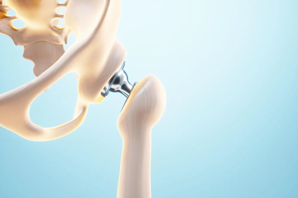
Hemiarthroplasty (Hemi Replacement of the Hip) is a surgical procedure where only one half of the hip joint is replaced. Specifically, the femoral head (the ball part of the ball-and-socket joint) is replaced with a prosthesis, while the acetabulum (the socket) is left intact. This procedure is commonly performed for certain types of hip fractures or severe arthritis.
This surgery helps relieve severe pain, restore hip function, and allows patients to regain mobility. It is especially beneficial for elderly patients or those with limited damage to the hip socket. With proper care, patients can return to daily activities more comfortably and safely.

Indications
Hemiarthroplasty is typically recommended when part of the hip joint is severely damaged but the socket remains healthy. The procedure helps reduce pain, restore function, and improve mobility, especially in elderly patients or those with limited hip damage.
- Femoral neck fractures (often in elderly patients with osteoporosis)
- Severe arthritis or degeneration limited to the femoral head
- Avascular necrosis of the femoral head
- Failed internal fixation of a hip fracture
Preoperative Preparation
Hemiarthroplasty is typically recommended when part of the hip joint is severely damaged but the socket remains healthy. The procedure helps reduce pain, restore function, and improve mobility, especially in elderly patients or those with limited hip damage.
Patient Evaluation
- Comprehensive medical history and physical examination
- Imaging studies: X-rays, MRI, or CT scans to assess the extent of damage
- Blood tests and other investigations to evaluate the patient’s fitness for surgery
Anesthesia
- General anesthesia or spinal/epidural anesthesia, depending on the patient’s health and preference
Patient Positioning
- The patient is placed in the supine or lateral decubitus position on the operating table
Surgical Procedure
Hemiarthroplasty involves replacing only the damaged half of the hip joint to relieve pain and restore function. The surgery is carefully performed to ensure precise placement of the prosthesis and a smooth recovery.
- Incision: A surgical incision is made on the lateral or posterior aspect of the hip. The choice of approach depends on the surgeon’s preference and the patient’s anatomy.
- Exposure: Muscles and soft tissues are carefully retracted to expose the hip joint. The hip is dislocated to access the femoral head.
- Femoral Head Removal: The femoral head is removed using surgical instruments, and the femoral canal is prepared for the prosthesis.
- Surgeons may use a variety of tools, including reamers and broaches, to shape the canal to fit the prosthesis snugly.
Prosthesis Insertion
The chosen prosthesis is inserted into the femoral canal.
Prostheses can be cemented or uncemented
- Cemented: Bone cement (polymethyl methacrylate) is used to fix the prosthesis in place.
- Uncemented: The prosthesis has a porous surface that allows bone to grow into it, securing it over time.
Reduction and Closure
- The hip is reduced back into its normal position.
- Muscles and soft tissues are reattached and sutured.
- The incision is closed in layers, and a sterile dressing is applied.
Postoperative Care
After hemiarthroplasty, proper care is essential to ensure healing, prevent complications, and restore mobility. Patients are closely monitored and guided through a structured recovery plan.
Immediate Postoperative Care
- Monitoring vital signs and managing pain with medications
- Early mobilization with the help of physical therapists, typically starting with weight-bearing as tolerated
Physical Therapy
- A tailored rehabilitation program to restore strength, range of motion, and functional mobility
- Use of assistive devices like walkers or crutches as needed
Wound Care
- Regular inspection and care of the surgical wound to prevent infection
- Removal of sutures or staples typically occurs around 10-14 days post-surgery
Follow-Up
- Regular follow-up visits to monitor healing, prosthesis position, and function
- Imaging studies are as needed to ensure proper placement and integration of the prosthesis
Potential Complications
Like any major surgery, hemiarthroplasty carries some risks that patients should be aware of. Understanding these helps in early recognition and management of complications. Prompt attention to warning signs can greatly improve outcomes.
- Infection: Possible at the surgical site
- Blood Clots: Risk of deep vein thrombosis (DVT) or pulmonary embolism (PE)
- Dislocation of the Prosthesis: The ball may slip out of the socket
- Prosthesis Loosening or Failure: Over time, the implant may become unstable
- Nerve or Blood Vessel Injury: Potential damage during surgery
- Leg Length Discrepancy: One leg may end up slightly shorter or longer
Hemiarthroplasty is a commonly performed procedure, especially in elderly patients with hip fractures. The surgery aims to relieve pain, restore function, and allow early mobilization.
Success depends on careful preoperative assessment, meticulous surgical technique, and comprehensive postoperative care, including physical therapy and regular follow-up. With the expertise of Dr. Manoj Pahukar in Nagpur, patients can achieve significant pain relief and improved quality of life.
Restore Your Hip Function with Hemi Replacement Surgery
Don’t let hip pain or limited mobility affect your daily life. Hemi replacement surgery in Nagpur can help relieve pain, improve function, and get you back to an active lifestyle.
Frequently Asked Questions
How long does a hemi replacement implant usually last?
Most hemi replacement implants can last 10-15 years or more, depending on the patient’s activity level, bone quality, and overall health.
When can I start climbing stairs after hemi replacement surgery?
With proper physiotherapy, many patients can begin climbing stairs with support within 2-3 weeks, though it may take longer for some depending on strength and balance.
Are there any long-term restrictions after hemi replacement?
Patients are usually advised to avoid high-impact activities like running or jumping. Low-impact exercises such as walking, swimming, or cycling are generally encouraged.
How soon can I drive after the surgery?
Driving is usually possible within 6-8 weeks, once strength, mobility, and reflexes return. Always consult your surgeon before resuming driving.
Can I sleep in any position after hemi replacement surgery?
Initially, patients may need to avoid crossing their legs or sleeping on the operated side. After recovery, most people can return to comfortable sleeping positions with few restrictions.
What role does physiotherapy play in recovery?
Physiotherapy is essential for regaining mobility, strengthening the muscles around the hip, and reducing stiffness. Skipping physiotherapy can delay recovery and affect long-term outcomes.
Will I need walking aids permanently?
Most patients use walkers or crutches for a few weeks. With progress in therapy, they transition to a cane, and many eventually walk independently without aids.
What signs should I watch for after surgery that may need medical attention?
Severe pain, swelling, redness, fever, or difficulty moving the hip may indicate complications and should be reported to your surgeon immediately.
